Research

MR imaging can be used to measure structural changes in the brains of individuals with multiple sclerosis and is essential for diagnosis, longitudinal monitoring, and therapy evaluation. The North American Imaging in Multiple Sclerosis Cooperative steering committee developed a uniform high-resolution 3T MR imaging protocol relevant to the quantification of cerebral lesions and atrophy and implemented it at 7 sites across the United States. To assess intersite variability in scan data, we imaged a volunteer with relapsing-remitting MS with a scan-rescan at each site.

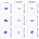
PREVAIL: In this work, our lab developed a model for predicting the degree and spatial pattern of MS lesion tissue recovery.

ABOUT PENNSIVE

The Penn Statistics in Imaging and Visualization Endeavor (PennSIVE) consists of a group of statisticians studying etiology and clinical practice through medical imaging. 

LOCATION

2nd Floor, Blockley Hall
University of Pennsylvania
423 Guardian Drive
Philadelphia, PA 19104-6021


SOCIAL MEDIA