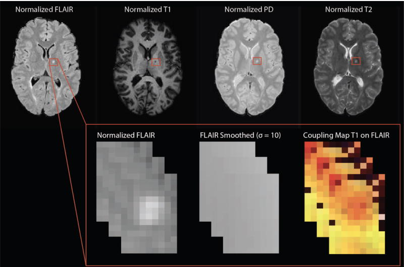
Magnetic resonance imaging (MRI) is crucial for in vivo detection and characterization of white matter lesions (WML) in multiple sclerosis. While WML have been studied for over two decades using MRI, automated segmentation remains challenging. Although the majority of statistical techniques for the automated segmentation of WML are based on single imaging modalities, recent advances have used multimodal techniques for identifying WML. Complementary modalities emphasize different tissue properties, which help identify interrelated features of lesions. MIMoSA, a fully automatic lesion segmentation algorithm which utilizes novel covariance features from inter-modal coupling regression in addition to mean structure to model the probability lesion is contained in each voxel, is proposed. Valcarcel et al, 2018.