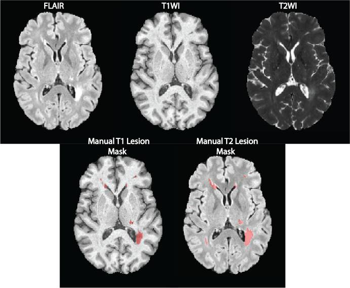
Magnetic resonance imaging (MRI) is crucial for in vivo detection and characterization of white matter lesions (WML) in multiple sclerosis (MS). The most widely established MRI outcome measure is the volume of hyperintense lesions on T2-weighted images (T2L). Unfortunately, T2L are non-specific for the level of tissue destruction and show a weak relationship to clinical status. Interest in lesions that appear hypointense on T1-weighted images (T1L) ("black holes") has grown because T1L provide more specificity for axonal loss and a closer link to neurologic disability. The technical difficulty of T1L segmentation has led investigators to rely on time-consuming manual assessments prone to inter- and intra-rater variability. Our group has develop edan automatic T1L segmentation approach, adapted from a T2L segmentation algorithm. Valcarcel et al. (2018)